
Robotic Treatment of Hiatal Hernia
Hiatal Hernia
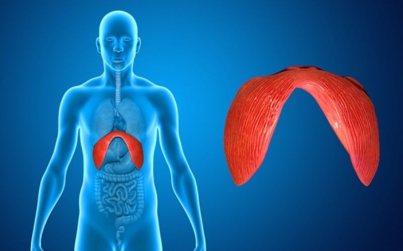
A hiatal hernia is the herniation (sliding) of a portion of the gastric fundus—sometimes small, sometimes large—into the thoracic cavity through the natural opening where the oesophagus descends into the abdomen.
It is a condition that is often diagnosed late.
It causes symptoms such as:
• Pain in the upper abdomen
• Regurgitation of food accompanied by burning, sometimes progressing to vomiting; symptoms worsen when bending forward. Patients often use multiple pillows during sleep.
Today, physicians investigate this condition primarily through upper gastrointestinal endoscopy.
Endoscopic findings may include:
• Presence of a hiatal hernia
• Oesophagitis of varying degrees due to reflux
• A precancerous condition (Barrett’s oesophagus)
When a hiatal hernia is diagnosed, and once conservative treatment options have been exhausted, if:
• Symptoms do not improve,
OR
• A precancerous condition such as Barrett’s oesophagus is present, the patient MUST undergo surgical treatment. A fundoplication must be performed—in other words, repositioning of the stomach into the abdomen and creation of a valve mechanism from the gastric fundus to prevent reflux (regurgitation) of gastric fluids and food into the oesophagus.
How?
Today, the operation is performed via the laparoscopic approach—the established LAPAROSCOPIC FUNDOPLICATION, most commonly the NISSEN technique.
BUT it may also be performed using robotic assistance—the ROBOTIC NISSEN FUNDOPLICATION.
The patient must be evaluated by a gastroenterologist.
The operation is performed under general anesthesia using highly advanced, state-of-the-art technological equipment and instruments.
Only four or five small skin incisions (5–10 mm) are required, through which the robotic fundoplication is carried out with the highest degree of SAFETY using the robotic system.
This technique requires specialized training of the surgeon and the operative team.
Advantages
The advantages of this technique include precise tissue dissection, minimal physiological burden on the patient, early mobilization, and rapid postoperative recovery.
The patient is discharged the following day, able to tolerate light food, and must follow a specific diet for 30–40 days.
If desired, the patient may return to work promptly (3–5 days) and resume usual activities.
The patient’s symptoms typically resolve quickly, and progression of Barrett’s oesophagus to potential oesophageal adenocarcinoma is prevented. (In such cases, ongoing follow-up by a gastroenterologist is essential.)